
Acid–base and the blood gas:
the stepwise read
Acid–base is the intern skill most often faked and least often taught properly — pattern-matched from the first two numbers, which works for the ordinary case and fails exactly when it matters. This hour is built the way the work actually arrives: a sequence to run every time, then the four disturbance patterns that account for most of what you will meet on the wards and in the unit, then the edge cases that defeat pattern-matching entirely. Interns rank acid–base among the topics they feel least prepared for (the evidence), and it is the one where a stepwise discipline pays off fastest.
Why this hour
Two habits produce most acid–base errors, and both are cured by the same discipline. The first is stopping early — reading the pH and the bicarbonate, naming “metabolic acidosis,” and moving on while a respiratory disorder and a second metabolic one sit unexamined in the same numbers. The second is reading the gas instead of the patient — treating a number rather than asking what process in this person’s body produced it. The sequence below fixes the first; the last step of the sequence fixes the second.
This page is a framework in the survival series’ house style, with one deliberate exception: acid–base cannot be taught without numbers, so the reference values and formulas live in the quick-reference figure and the arithmetic is worked in the eight cases below. No treatment thresholds, doses, or protocol steps appear anywhere on the page — those remain your faculty’s to teach from current references and institutional protocol, on a date that shows.
What interns leave able to do
- Run the full sequence aloud on any gas, in order, without skipping to the answer.
- Judge compensation against its expectation — and recognize that a compensation which does not fit means a second disorder, not an unusual patient.1
- Calculate and use the anion gap, and interrogate it further when the history does not explain it.
- Recognize the four patterns that account for most ward and unit gases — and name the causes that actually produce each in this hospital’s patients.
- Work the edge cases that defeat pattern-matching, up to and including a triple disturbance.
- Convert every finding into a differential and a first move: what process did this, and what happens in the next hour.
The sequence — the whole method
The order is the technique. Faculty supply the numbers behind each step from current references; what the intern owns is the discipline of never skipping one.
- Start with the patient and the clinical context. Who is this, what happened, and what do you already expect the gas to show? A prediction made before the numbers is the fastest way to learn.
- Is the patient acidemic or alkalemic? The pH names the dominant process — and only the dominant one.
- Which system is driving it — respiratory or metabolic? Read the carbon dioxide and the bicarbonate together, and say which one moves with the pH.
- Is compensation appropriate? Compare the observed compensation against its expected value.1 This is the step interns skip and the step that finds second disorders: compensation that overshoots or falls short is not an eccentric patient — it is another disturbance announcing itself.
- Calculate the anion gap — every time, including when the bicarbonate looks normal, because a normal bicarbonate can hide an acidosis and an alkalosis canceling each other. Adjust it for the albumin: albumin is the largest unmeasured anion, so a low albumin lowers the calculated gap and can hide a raised-gap acidosis outright.2 The physiology is not in dispute; the exact coefficient is debated, so calculate the correction and hold it as a prompt to look harder rather than as a precise second number.
- If the gap is raised, compare its size to the change in bicarbonate — the delta comparison, which reveals whether a second metabolic process rides alongside the gap acidosis.
- Return to the patient. Name the disorders, then the process behind each — and the differential the physiology supports.2 A gas read without a differential is arithmetic, not medicine.
- Say what you will do in the next hour — the treatment follows the process, not the number, and the intern’s job is the first move and the right call.
The quick-reference guide
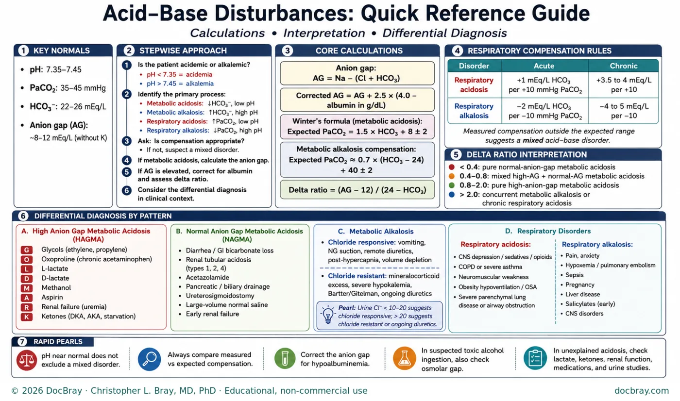
One card carries the whole method: the normals, the stepwise approach, the calculations, the compensation expectations, the delta-ratio bands, the differential by pattern, and the pearls. Print it, photograph it, keep it where you read gases — and use the rest of this page to learn what it cannot teach, which is judgment.
What you will actually meet
Four patterns account for the large majority of gases an internal-medicine resident reads. Learn where each one lives, what actually causes it in this hospital’s patients, and what the intern does next — the differential lists on the card become useful only once they are attached to the wards they come from.
1. High anion gap metabolic acidosis — the sick-patient pattern. The one you will meet most often in the emergency department and the unit, and the one that most often means something urgent. In practice the gap is nearly always lactate, ketones, kidney failure, or an ingestion — and the mnemonic on the card exists to stop you forgetting the rarer members of that list. The intern’s move: a raised gap is not a diagnosis, it is a prompt — which anion, and why? Send what identifies it, treat the process rather than the pH, and remember that lactate has many causes besides sepsis, several of which are not hypoperfusion at all.
2. Normal anion gap metabolic acidosis — the quieter one. Less dramatic, frequently iatrogenic, and easy to attribute to nothing. It shows up in the patient with high-volume diarrhea or a high-output ostomy, in the patient with a renal tubular acidosis, in drainage losses, and after large-volume saline resuscitation — which makes it, on some services, something the team caused. The intern’s move: separate gastrointestinal loss from renal loss, because the workups diverge there; the card’s urine studies are how that question gets answered.
3. Metabolic alkalosis — the ward’s regular. Common, usually unglamorous, and usually explained by the chart before any test: vomiting, nasogastric suction, diuretics, volume depletion. The important split is chloride-responsive versus chloride-resistant, because the first improves with the volume and chloride the patient is missing while the second will not, and pursuing it as though it were the first wastes days. The intern’s move: read the chart for losses and drugs first, then use the urine chloride the card points you to before deciding which kind you are looking at.
4. Respiratory disorders — where acute and chronic are different diseases. Respiratory acidosis in the patient with obstructive lung disease, sedation, neuromuscular weakness, or obesity hypoventilation; respiratory alkalosis in pain, anxiety, hypoxemia, pulmonary embolism, sepsis, pregnancy, liver disease, or early salicylate toxicity. The compensation table on the card matters here more than anywhere: the same carbon dioxide value means something entirely different in the patient who has lived with it for years than in the one who arrived that way this afternoon. The intern’s move: decide acute versus chronic before you decide anything else — that single question drives whether this is a chart finding or a call for help (the ICU hour’s hypercapnic-failure teaching is the escalation half).
Two things about the differential lists on the card are worth saying out loud in the room. Frequency is local: which causes you actually meet depends on your hospital’s population — a liver service, a dialysis unit, and a rural emergency department produce different acid–base practices. And the list is a prompt, not a protocol: its job is to stop you settling on the first plausible cause when a second one is sitting in the same chart.
Eight gases to work
Four ordinary, four that break pattern-matching. Read the numbers, run the sequence out loud, commit to an answer — then open the read. Albumin is given because the sequence requires it, and every set is internally consistent, so the arithmetic on the card will reproduce these answers exactly.
1. The 24-year-old with vomiting and thirst. Type 1 diabetes, insulin pump failed two days ago. Breathing deeply, dry mucous membranes, alert.
ABG pH 7.22 · PaCO2 20 · HCO3− 8
BMP Na 132 · K 5.4 · Cl 95 · HCO3− 8 · BUN 32 · Cr 1.4 · glucose 480 · albumin 4.0
Show the read
Acidemic (pH 7.22), and the bicarbonate moves with the pH — a metabolic acidosis is the primary process.
Compensation: Winter’s predicts a PaCO2 of 1.5 × 8 + 8 = 20 ± 2. Measured 20 — appropriate. That deep breathing is the compensation working, not a second disorder.
Gap: 132 − (95 + 8) = 29, raised. Albumin is normal, so no correction. Delta ratio = (29 − 12) / (24 − 8) = 1.06 — between 0.8 and 2.0, so a pure high-gap acidosis with nothing else underneath.
Answer: a simple high-anion-gap metabolic acidosis with appropriate respiratory compensation — diabetic ketoacidosis. The teaching point: the potassium reads high while total-body potassium is severely depleted — insulin will drive it into cells quickly, so the number falls once treatment starts. That is why potassium is followed closely from the outset, why repletion begins well before the number looks low, and why a low initial potassium changes the order of treatment entirely. The thresholds that govern all of that live in the glycemic-emergencies hour and your own protocol — do not infer them from this case.
2. The 58-year-old with a week of diarrhea. Ten watery stools a day since a course of antibiotics. Tired, thirsty, no abdominal pain.
ABG pH 7.32 · PaCO2 30 · HCO3− 15
BMP Na 138 · K 3.1 · Cl 114 · HCO3− 15 · BUN 18 · Cr 1.0 · glucose 95 · albumin 4.0
Show the read
Acidemic, bicarbonate low and moving with the pH — metabolic acidosis.
Compensation: Winter’s predicts 1.5 × 15 + 8 = 30.5 ± 2. Measured 30 — appropriate.
Gap: 138 − (114 + 15) = 9 — normal. Note the chloride of 114 rising as the bicarbonate falls: this is a hyperchloremic, normal-anion-gap acidosis, and no delta ratio is needed because there is no gap to apportion.
Answer: normal-anion-gap metabolic acidosis from gastrointestinal bicarbonate loss, with the hypokalemia that travels with high-volume diarrhea. The teaching point: the next question separates gastrointestinal from renal loss — urine studies answer it, and a renal tubular acidosis is the alternative that changes the workup entirely. Also ask what the antibiotics started.
3. The 70-year-old with four days of nasogastric suction. Post-operative ileus, tube draining well, minimal intake, on no diuretic.
ABG pH 7.53 · PaCO2 50 · HCO3− 40
BMP Na 140 · K 2.9 · Cl 88 · HCO3− 40 · BUN 40 · Cr 1.3 · albumin 4.0 · urine Cl− 8
Show the read
Alkalemic (pH 7.53) with a high bicarbonate moving with the pH — metabolic alkalosis.
Compensation: expected PaCO2 ≈ 0.7 × (40 − 24) + 40 = 51 ± 2. Measured 50 — appropriate hypoventilation, not a second disorder. Treat that band generously: the respiratory response in metabolic alkalosis is the least reliable of the compensation relationships, because hypoxemic drive limits how far a patient will hypoventilate, and wider tolerances are commonly quoted. Gap 140 − (88 + 40) = 12, normal.
Answer: a chloride-responsive metabolic alkalosis from gastric losses — the urine chloride of 8 is the confirming test, and it is the number that decides the treatment. The teaching point: this alkalosis is sustained by what the patient is missing (chloride, volume, potassium), so it corrects when those are replaced. Contrast it with the chloride-resistant kind — a urine chloride comfortably above 20 in a patient who is not on diuretics — which will not budge with saline and points instead toward mineralocorticoid excess or severe potassium depletion.
4. The 66-year-old with COPD, seen in clinic. Baseline breathlessness, no worse than usual, here for a routine visit. Comfortable, speaking full sentences.
ABG pH 7.32 · PaCO2 68 · HCO3− 34
BMP Na 140 · K 4.2 · Cl 96 · HCO3− 34 · BUN 16 · Cr 0.9 · albumin 4.0
Show the read
Acidemic, and here the carbon dioxide moves with the pH — a respiratory acidosis is primary.
Acute or chronic? The whole read turns on this. The PaCO2 is 28 above 40; an acute rise would lift the bicarbonate by about 1 per 10, to roughly 27. A chronic rise lifts it by 3.5 to 4 per 10 — to roughly 34 to 35. Measured 34: chronic, with appropriate metabolic compensation. Gap 140 − (96 + 34) = 10, normal.
Answer: chronic respiratory acidosis with appropriate metabolic compensation — this is his baseline. Note that the same carbon dioxide of 68 arriving acutely in a previously well patient would be an emergency, and it would look different: the bicarbonate would sit near 27, not 34, because the kidney has not had time to respond. The teaching point: treat the patient, not the gas. Reacting to the number in a stable outpatient is how harm happens here — and note that aggressive oxygen would raise this carbon dioxide rather than lower it. Escalation criteria for the acute version live in the ICU hour.
5. The 61-year-old with cirrhosis and fever. Admitted with ascites and hypotension responding to fluids. The intern reports “gas is basically fine, no gap.”
ABG pH 7.36 · PaCO2 37 · HCO3− 20
BMP Na 136 · K 4.0 · Cl 106 · HCO3− 20 · BUN 22 · Cr 1.2 · albumin 1.8 · lactate 4.2
Show the read
The reported read is wrong, and the albumin is why. Raw gap: 136 − (106 + 20) = 10 — which does look normal.
Corrected for albumin: 10 + 2.5 × (4.0 − 1.8) = 15.5 — raised. Albumin is the largest unmeasured anion, so a low albumin lowers the calculated gap and can hide a real acidosis outright.2 The lactate of 4.2 is the anion the corrected gap is pointing at.
Compensation: Winter’s predicts 1.5 × 20 + 8 = 38 ± 2; measured 37 — appropriate. Delta ratio using the corrected gap = (15.5 − 12) / (24 − 20) = 0.88, consistent with a pure high-gap process — though note how fragile that number is when both terms are small: a shift of one in the sodium, chloride, or bicarbonate moves it enough to change the band, which is why the ratio informs the read rather than settling it.
Answer: a hidden high-anion-gap lactic acidosis in a hypoalbuminemic patient — and in a febrile, hypotensive cirrhotic, that is a sepsis workup, not a reassuring gas. The teaching point: the patients in whom the gap hides are exactly the patients in whom it matters most. Correcting for albumin is part of calculating the gap, not a refinement.
6. The 45-year-old with diabetes, vomiting for three days. Poor intake, taking a thiazide for hypertension. Ketones present.
ABG pH 7.34 · PaCO2 38 · HCO3− 20
BMP Na 138 · K 3.4 · Cl 88 · HCO3− 20 · BUN 30 · Cr 1.3 · glucose 420 · albumin 4.0
Show the read
Mildly acidemic with a bicarbonate of 20 — a picture that looks unimpressive until you calculate.
Gap: 138 − (88 + 20) = 30. That is a severe gap acidosis sitting behind an almost ordinary bicarbonate.
Delta ratio: (30 − 12) / (24 − 20) = 4.5. Well above 2, which means the bicarbonate has not fallen anywhere near as far as the gap has risen — something is holding it up. That something is a concurrent metabolic alkalosis from three days of vomiting and the thiazide.
Compensation: Winter’s predicts 38 ± 2 for a bicarbonate of 20; measured 38 — appropriate, so the respiratory system is not a third player here.
Answer: a high-anion-gap ketoacidosis plus a metabolic alkalosis. The teaching point: the delta ratio is what makes the second disorder visible, and the clinical consequence is real — treat this patient as though the bicarbonate of 20 reflected mild illness and you will underestimate a ketoacidosis that needs full treatment.
7. The 58-year-old cirrhotic admitted with pneumonia. Febrile, tachypneic, vomiting for two days before arrival, hypotensive on arrival.
ABG pH 7.52 · PaCO2 28 · HCO3− 22
BMP Na 140 · K 3.2 · Cl 92 · HCO3− 22 · BUN 35 · Cr 1.8 · albumin 3.0 · lactate 5.0
Show the read
Alkalemic (pH 7.52) with a low carbon dioxide — so the primary process reads as a respiratory alkalosis, and an intern who stops here reports “alkalosis from sepsis” and moves on. Three disorders are present.
Gap: 140 − (92 + 22) = 26, corrected for albumin = 26 + 2.5 × (4.0 − 3.0) = 28.5 — a severe high-gap metabolic acidosis, entirely invisible in the pH. Note that the lactate of 5.0 does not account for all of it: the ketoanions of two days without intake and the kidney injury are contributing, and asking which anions make up this gap is the habit the case is teaching.
Delta ratio: (28.5 − 12) / (24 − 22) = 8.25 — far above 2, so a metabolic alkalosis is also present, from the vomiting.
Compensation check: for a bicarbonate of 22, Winter’s predicts a PaCO2 of 41 ± 2. Measured 28 — far lower than expected, confirming the respiratory alkalosis as its own disorder rather than compensation.
Answer: a triple disturbance — high-gap lactic acidosis, metabolic alkalosis, and respiratory alkalosis — in a patient whose story explains all three. The teaching point: the alkalemic pH is the trap. Two of these three disorders were about to be missed, and the one that matters most tonight is the one the pH concealed.
8. The 31-year-old brought in confused, with tinnitus. Found at home; an empty bottle of an over-the-counter analgesic nearby. Hyperventilating, febrile, diaphoretic.
ABG pH 7.43 · PaCO2 22 · HCO3− 14
BMP Na 140 · K 3.6 · Cl 102 · HCO3− 14 · BUN 14 · Cr 1.0 · glucose 88 · albumin 4.0
Show the read
The pH is nearly normal — 7.43 — and two significant disorders are present underneath it.
Gap: 140 − (102 + 14) = 24, clearly raised: a high-anion-gap metabolic acidosis. Delta ratio = (24 − 12) / (24 − 14) = 1.2, consistent with a pure gap process.
Compensation: Winter’s predicts 1.5 × 14 + 8 = 29 ± 2. Measured 22 — well below the expected range, so this is not compensation. A primary respiratory alkalosis is present as well.
Answer: the simultaneous respiratory alkalosis and high-gap metabolic acidosis characteristic of salicylate toxicity — the drug stimulates the respiratory center directly while generating the acidosis. The teaching point: a normal pH excludes nothing, and this combination in a confused patient should prompt a salicylate level and an urgent call to the poison center and nephrology, because the treatment thresholds and the dialysis decision are time-critical and belong to them. One recognition point worth carrying: the hyperventilation here is protective — anything that blunts it, sedation and intubation among them, can be catastrophic in this poisoning, which is why the airway decision is never a reflex.
Every case here is a fictional composite constructed for teaching, with internally consistent chemistry; values are illustrative and not reference standards. Verify the formulas and expectations against current references before teaching, and treat management as belonging to the supervised team under institutional protocol.
The edge cases — where pattern-matching fails
These are the gases that separate a resident who reads numbers from one who reads patients. Each deserves a worked example; a program that builds them once can reuse them every year.
The normal-looking gas. A near-normal pH with an unremarkable bicarbonate, hiding two opposing disorders that happen to cancel. Found only by someone who calculated the gap when nothing appeared to be wrong — which is why the sequence calculates it every time.
The gap in the hypoalbuminemic patient. The commonest reason a raised-gap acidosis is missed outright: albumin is the largest unmeasured anion, so a low albumin lowers the calculated gap and can make a significant acidosis read as normal.2 It hides most reliably in exactly the critically ill patient in whom it matters most.
The delta-ratio surprise. A raised gap that is not the whole story: the ratio of the gap’s rise to the bicarbonate’s fall reveals a second metabolic process riding alongside — a normal-gap acidosis underneath, or a metabolic alkalosis on top. This is the step that most often changes the plan, and the one interns skip.
Compensation mistaken for a second disorder, and the reverse. Expected compensation has a range, not a value.1 Judging it by feel produces both errors: a normal patient over-diagnosed with a mixed picture, and a genuine second disorder waved through as “appropriate compensation.”
The chloride-resistant alkalosis. The metabolic alkalosis that does not improve with volume and chloride, because its cause is mineralocorticoid excess, severe potassium depletion, ongoing diuretics, or one of the inherited tubulopathies. The tell is on the card; the trap is treating every alkalosis as though it were the common kind.
The unexplained gap — and the ingestion you have not considered. When the history does not account for the gap, widen the differential rather than assume the usual cause: the card’s pearl about checking an osmolar gap in suspected toxic-alcohol ingestion exists because that diagnosis is time-critical and easy to miss, and because the specific antidotes and dialysis decisions belong to the toxicologists and nephrologists you should already be calling.
The triple disturbance. The capstone: three simultaneous disorders — classically a raised-gap acidosis with a metabolic alkalosis riding on it and a respiratory disorder on top — in a patient whose story explains all three (the vomiting, the sepsis, the lung disease, the diuretic). Deliberately impossible to pattern-match: the pH may be nearly normal and the bicarbonate unremarkable, with the whole picture visible only to someone who calculated the gap when nothing looked wrong and then compared the delta. How to run it: give the room the gas and the story, let them commit before any arithmetic, then walk the sequence and watch the second and third disorders appear at steps four and six. The teaching lands in the distance between the confident first answer and the complete one.
The gas that does not fit the patient at all. Sampling, timing, and transport errors are real; so is a venous sample labeled arterial. Repeating a discordant gas before acting on it is judgment, not indecision — and knowing what a venous sample can and cannot answer prevents both the unnecessary arterial stick and the wrong conclusion from an easy one (the procedure lab teaches the stick itself).
And the error that outranks all of them: treating the number. The bicarbonate is not the disease and the pH is not the patient. Every teaching gas in this hour ends the same way — with the process named, the differential stated, and the next hour’s actions said out loud.
Running the hour
| Minutes | Block |
|---|---|
| 0–5 | Frame: “pattern-matching works until it doesn’t — today you get a sequence, the four patterns, and the cases that break them” |
| 5–15 | The sequence on the board, built with the room; the quick-reference guide handed out and walked once |
| 15–32 | The four common patterns, one worked gas each — run aloud by a different intern, with the causes named from your own service’s patients |
| 32–50 | Edge cases: the hypoalbuminemic gap and a delta-ratio surprise, then the triple disturbance — commit first, then walk the sequence and find all three |
| 50–60 | Each gas revisited for its differential and first move · pocket card |
Watch for, and debrief by name: the intern who announces the answer from the first two numbers — correct, and still the habit being retired; the gap skipped when the bicarbonate looks normal; the albumin never mentioned; compensation judged by feel rather than expectation; the alkalosis assumed to be the common kind; and the confident triple-disturbance answer that names only the loudest disorder. Bring gases from your own patients where de-identification allows — the sequence teaches best on the hospital’s real numbers, and the common-pattern block is where local case mix should replace the generic list.
Pocket card
- Patient first: what do you expect this gas to show?
- Acidemic or alkalemic → which system is driving it.
- Check compensation against expectation. Doesn’t fit = a second disorder.
- Calculate the gap every time — even when the bicarbonate looks fine — and correct it for albumin.
- Gap raised? Compare the delta — that’s where the third disorder hides.
- Alkalosis: read the chart for losses and drugs, then ask if it’s chloride-responsive.
- Respiratory: acute or chronic? Decide that before anything else.
- Name the process, not the number. Then say your next hour.
Notes
Three cautions worth passing to faculty before this hour runs. The compensation relationships are approximations from old and small studies, and the respiratory response in metabolic alkalosis is the least predictable of them — teach every expectation as a range with a purpose, not as a law.2 Substitute your own laboratory’s normal anion gap for the value used in the worked cases: modern analyzers put it lower than the traditional figure, and both the gap and every delta ratio derived from it shift with that baseline. And the delta ratio’s denominator is contaminated whenever a respiratory disorder coexists, because the respiratory process moves the bicarbonate itself — in the mixed cases below it still points the right direction, but it is a signpost rather than a measurement.
This hour runs in the weeks-2–8 survival series, one conference hour, after the data stations have taught the systematic read — this is the level deeper: from the identified disorder to its differential and first response. Faculty bring their own worked gases; the edge cases are worth building once and reusing every year. The quick-reference guide is free to print and hand out for teaching under the terms in the site footer.
This page is a curriculum framework, not clinical instruction. Its prose states no thresholds or treatment targets; the reference values, formulas, compensation expectations, and differential lists appear only inside the quick-reference figure, which is a teaching aid rather than a standard of care — verify its values and formulas against current references before teaching from it, and treat institutional protocols as governing. Management belongs to the supervised team. Last reviewed July 2026.
Sources
- Albert, M. S., Dell, R. B., & Winters, R. W. (1967). Quantitative displacement of acid–base equilibrium in metabolic acidosis. Annals of Internal Medicine, 66(2), 312–322. https://pubmed.ncbi.nlm.nih.gov/6016545/ The source of the expected-respiratory-compensation relationship still taught at the bedside; the point of citing it is that compensation has a measurable expectation, not that a formula should be memorized without one. ↩1 ↩2 ↩3
- Berend, K., de Vries, A. P. J., & Gans, R. O. B. (2014). Physiological approach to assessment of acid–base disturbances. New England Journal of Medicine, 371(15), 1434–1445. https://pubmed.ncbi.nlm.nih.gov/25295502/ ↩1 ↩2 ↩3 ↩4 ↩5